
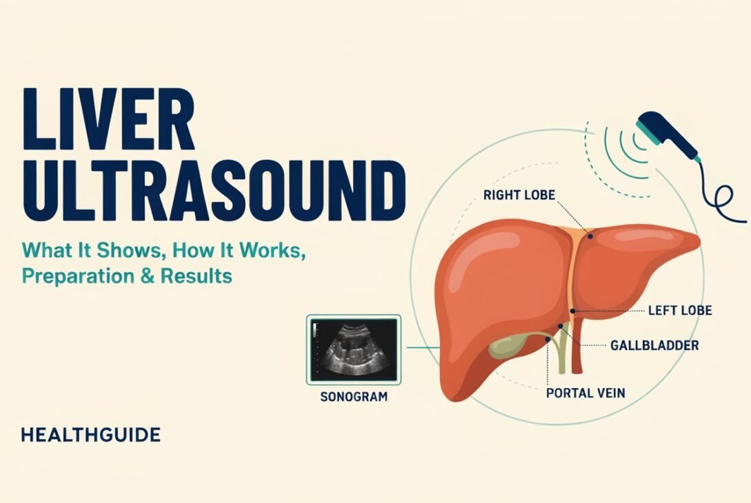
When a doctor utters the words “liver ultrasound,” it’s only reasonable to feel a bit nervous. Most people think of the machines they’ve seen in the movies or on TV shows regarding pregnancy, not realizing this same technology plays a vital role in checking the health of one of the body’s hardest working organs.
A liver ultrasound is one of the simplest, safest and most informative procedures that can be performed to check on the condition of your liver. No needles, no radiation, no hospital stay. But it can tell you a surprising amount about what’s going on within your body.
This guide gives you the lowdown in basic, straightforward language on everything you need to know: what the test shows, how it truly works, how to prepare and how to make sense of your results.
What Is an Ultrasound of the Liver?
Liver ultrasonography is a painless imaging procedure that uses high-frequency sound waves to produce real-time images of the liver and surrounding structures. It is sometimes called a hepatic ultrasound or abdominal ultrasound when surrounding organs such as the gallbladder, bile ducts, and spleen are involved.
X-ray and CT scans use ionizing radiation, which is fully avoided in ultrasound. This makes it the imaging modality of choice for many liver diseases, particularly when serial monitoring over time is required.
In simple terms, the equipment emits sound waves into your body. Each tissue type reacts to these waves differently, sending back a unique echo pattern for the machine to read. A computer turns these echoes into a live picture on a screen so the doctor or sonographer can view the size, shape, texture and blood flow of the liver.
Why Doctors Order a Liver Ultrasound
Doctors don’t order this test randomly. In most cases, something in your symptoms or test results points the doctor toward this exact test. Common triggers include:
- Abnormal liver function test (LFT) results in blood work
- Ongoing discomfort near the upper right quadrant of the belly, with no clear cause
- Jaundice (yellowing of skin or eyes)
- Unexplained fatigue or weight loss
- Swelling or bloating in the abdomen
- History of hepatitis B or C
- Chronic alcohol use
- Monitoring known liver disease, such as fatty liver or cirrhosis
- Family history of liver conditions
- Suspected gallstones or bile duct blockage
In many cases, this test is the starting point. If something unusual shows up, your doctor may order additional imaging like a CT scan, MRI, or a liver biopsy for more detail.
What Is Liver Ultrasound Showing?
This is where most people are most curious. This is what the test can identify or assist you assess:
1. Size and Shape of the Liver
A big liver (hepatomegaly) may be an indication of infection, fatty liver disease, or other underlying disorders. Ultrasound provides a clear image of the whole liver size.
2. Steatosis
One of the most common finds nowadays. The more fat there is in the liver tissue, the brighter and denser the appearance will be on the scan – sometimes referred to as enhanced echogenicity.
3. Fibrosis and Cirrhosis
Chronic liver injury may result in a rough or nodular surface and interior texture of the liver. Ultrasound can detect early structural alterations.
4. Cysts of the liver
The fluid-filled sacs are normally innocuous and are sometimes spotted unintentionally on a scan.
5. Tumors or Masses
Benign growths (such as hemangiomas) and worrisome masses may be discovered. More tests are often needed to find out if anything is malignant.
6. Bile duct obstruction
If bile can not flow normally, a major clue for gallstones or tumors pressing on the duct system is dilated ducts on the scan.
7. Circulation problems
Doppler ultrasound is a unique type of ultrasound that measures the flow of blood through the veins and arteries of the liver. This test helps to find blood clots or abnormal blood pressure in the portal vein (portal hypertension).
Types of Liver Ultrasound
Not all liver scans are created equal. Depending on your symptoms, your doctor may order one of these variations:
- Standard abdominal ultrasonography — broad survey of the liver and surrounding organs
Doppler ultrasonography – direction and speed of blood flow
Elastography (FibroScan or shear-wave elastography) — evaluates the stiffness of the liver to assess fibrosis or cirrhosis without a biopsy
Contrast-enhanced ultrasonography — employs a particular contrast agent to better characterize liver lesions.
Each type has a particular purpose for diagnosis and your doctor will choose based on what you are concerned about.
What Is a Liver Ultrasound?
It’s easier than most people think.
- Step-By-Step Guide
You’re lying on an exam table, generally on your back, but sometimes on your side.
A gel is applied to your upper abdomen. This gel assists the sound waves to move smoothly without air gaps interfering with the image.
A handheld device called a transducer is softly put against your skin and moved over your abdomen.
Your liver tissue reflects sound waves and a computer instantaneously turns them into moving visuals on a monitor.
The sonographer takes pictures from several directions, sometimes asking you to hold your breath for a few seconds to get better shots.
And when you’re done, you wipe off the gel.”
Usually the complete scan takes half an hour or less from beginning to end. It does not hurt . You may feel some pressure from the transducer but that is all .
How to Prepare for a Liver Ultrasound
Preparation is simple but important, since food and gas in the digestive tract can interfere with image clarity.
Common Preparation Guidelines
- Fast for 6–8 hours before the test. Food, especially fatty meals, can cause the gallbladder to contract and gas to build up, both of which reduce image quality.
- Avoid chewing gum or smoking before the scan, as these can increase swallowed air.
- Drink water only if instructed. Some clinics ask for a full bladder if other organs are being checked simultaneously; others don’t require this for liver-focused scans.
- Because the technician needs direct access to your bare skin, choose an outfit that’s easy to adjust in the moment
- Inform your doctor of medications you’re taking, though most don’t need to be stopped for this test.
Always follow the specific instructions given by your healthcare provider or the imaging center, since protocols can vary slightly.
What To Expect When You Take The Test
Most patients report the encounter as uneventful. You will feel the coolness of the gel and a light pressure as the technician moves the transducer. No needles, no radiation, no down time.
In general you can:
- Then drive home yourself
Eat normally immediately after the scan
Return to normal work or regular activities straight away
If you’re having an elastography scan to assess liver stiffness, the method is almost same, with additional readings recorded to assess tissue elasticity
Understanding Your Liver Ultrasound Results
Results are usually reviewed by a radiologist, who prepares a report for your referring doctor. Here’s how to interpret common findings.
Normal Results
A healthy liver typically shows:
- Smooth, well-defined edges
- Uniform texture throughout
- Normal size for your body dimensions
- No visible masses, cysts, or fluid buildup
- Normal blood flow patterns
Abnormal Results
Depending on what’s found, your report might mention:
| Finding | Possible Meaning |
|---|---|
| Increased echogenicity | Fatty liver disease |
| Nodular surface | Cirrhosis or advanced scarring |
| Dilated bile ducts | Blockage, possibly from stones or tumor |
| Focal lesion or mass | Cyst, benign tumor, or requires further testing |
| Enlarged liver | Inflammation, infection, or fat accumulation |
It’s important to remember that an abnormal ultrasound doesn’t automatically mean cancer or severe disease. Many findings, like small cysts or mild fatty changes, are common and not dangerous. Your doctor will interpret results alongside your blood tests, symptoms, and medical history.
Liver Ultrasound vs Other Liver Tests
People often ask how ultrasonography compares to other diagnostic modalities.
- Ultrasound vs. CT scan: Ultrasound is radiation-free and less costly, and is good for follow-up. CT scans provide more detail but use radiation.
Ultrasound vs. MRI: MRI delivers the best soft-tissue detail, but it is more expensive and takes longer. Ultrasound is quicker and more convenient.
Ultrasound vs. liver biopsy: Biopsy is still the gold standard for detecting some liver illnesses on a cellular level, but it’s an intrusive procedure. Ultrasound elastography is now frequently used as a non–invasive method to measure fibrosis.
Ultrasound is the first step in most clinical routes with more advanced testing reserved for cases that need further clarification.
Is a Liver Ultrasound Safe?
Yes. This is one of the safest imaging tests in modern medicine. It uses sound waves, not radiation, and has no known harmful side effects. It’s even considered safe during pregnancy, which is part of why ultrasound technology is so widely trusted across medical specialties.
Questions Frequently Asked
1. Is a liver ultrasound painful?
Nope. You may feel some pressure from the transducer, but the test is painless.
2. How long does liver ultrasound take?
Usually 15 to 30 minutes depending on how many organs are being looked at.
3. Is liver cancer visible on a liver ultrasound?
It can find worrisome lumps and is usually followed up with other testing, such as a CT scan, MRI or biopsy, to confirm a cancer diagnosis.
4. Do I need to fast for a liver ultrasound?
Many clinics ask you to fast for 6-8 hours prior to the scan to increase the clarity of the images, especially for imaging of the gallbladder and bile ducts.
5. Can ultrasonography be used to identify fatty liver disease?
Yes. Ultrasound is often the first-line method to identify fatty liver, which is typically characterized by increased brightness (echogenicity) on the scan.
6. How reliable is a liver ultrasound?
It is quite good at picking up structural abnormalities, fatty liver, cysts and blockages, although very small lesions or early disease may need more sensitivity imaging.
7.When can I expect to receive the results?
This depends on the facility, but most patients get findings in a few days. Sometimes it’s faster if the case is urgent.
Conclusions.
A liver ultrasound is a rapid, pain-free test that shows the health of your liver without using radiation. It can be used to diagnose abnormal blood work, to track a known ailment, or merely to rule out concerns. This test provides important knowledge without the hazards of more intrusive treatments.
So if your doctor has recommended you have an ultrasound of your liver, don’t panic. It requires little preparation and is a quick operation, and the information it collects can be critical in maintaining your long-term health. As usual, talk over your exact results and what to do next with your health care provider, who knows the whole story of your medical history and can help point you in the right direction