
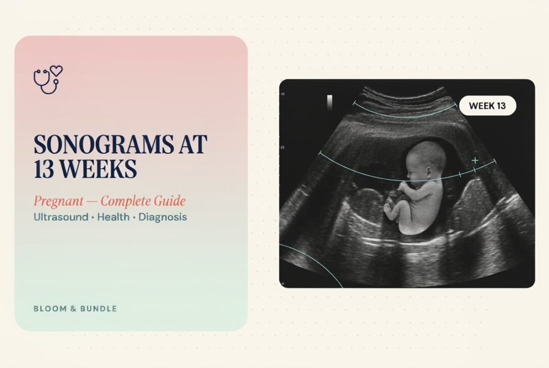
Reaching 13 weeks is a milestone many expecting parents look forward to. It marks the end of the first trimester and often comes with a scheduled ultrasound. If you have a sonogram coming up at this stage, you probably have questions about what the scan shows, how your baby is developing, and what to expect during the appointment.
This guide walks through everything relevant to sonograms at 13 weeks pregnant — what the ultrasound looks for, how it compares to earlier scans, fetal size and development, and when you might learn the baby’s sex. As always, this article is for general information only and doesn’t replace advice from your OB-GYN or midwife.
What Is a Sonogram at 13 Weeks Pregnant?
A sonogram at 13 weeks pregnant is an ultrasound scan performed near the end of the first trimester. It uses sound waves to create real-time images of the fetus, placenta, and surrounding structures inside the uterus.
At this stage, the scan is often combined with what’s known as the nuchal translucency (NT) screening, which measures the fluid at the back of the baby’s neck. This measurement, along with maternal blood tests, helps assess the likelihood of certain chromosomal conditions, such as Down syndrome.
Why This Scan Matters
- Confirms the pregnancy is progressing on schedule
- Checks fetal heartbeat and movement
- Measures the baby for accurate due date confirmation
- Screens for early signs of chromosomal or structural concerns
- Assesses the number of fetuses (in case of multiples)
Doctors typically consider this one of the more informative scans of the first trimester because the fetus is large enough to observe key structures clearly.
Size of a 13 Week Fetus
One of the most common questions parents ask is how big their baby is at this point. The size of a 13 week fetus is often compared to a fruit for easy visualization.
At 13 weeks, the average fetus measures:
- Length (crown to rump): approximately 2.9 to 3.1 inches (about 7.4 cm)
- Weight: roughly 0.7 to 0.8 ounces (around 23 grams)
- Comparable size: about the size of a peach or a lemon
By this week, most of the baby’s organs have formed and are beginning to function, though they will continue maturing throughout the pregnancy. Fingers and toes are fully separated, vocal cords are starting to develop, and the baby can make small movements, even though these usually aren’t felt by the mother yet.
What the Ultrasound Can Show at This Stage
During a 13-week sonogram, the sonographer typically checks:
- Fetal heart rate (usually between 140–170 beats per minute)
- Head, abdomen, and limb measurements
- Nuchal translucency thickness
- Presence and position of the placenta
- Amniotic fluid levels
- Basic anatomical structures, including the skull, spine, and limbs
While some structures are visible, a more detailed anatomy scan is usually performed later, around 18 to 22 weeks, when organs are larger and easier to evaluate.
How Does the 13-Week Scan Compare to Earlier Ultrasounds?
Understanding how this scan fits into the overall prenatal ultrasound schedule helps set realistic expectations.
8 Week Ultrasound
An 8 week ultrasound is often the first scan performed, usually to confirm the pregnancy and check for a heartbeat. At this stage:
- The embryo measures about 0.6 to 0.7 inches long
- A heartbeat is often detectable, typically ranging from 150–170 bpm
- The scan confirms the pregnancy is located in the uterus (ruling out ectopic pregnancy)
- Gestational age and due date are estimated based on measurements
This early scan is usually performed transvaginally, since the embryo is still very small and harder to visualize through the abdomen.
12 Week Ultrasound
The 12 week ultrasound is very similar in purpose to the 13-week scan and is often used interchangeably, depending on the clinic’s scheduling. This scan usually includes:
- Nuchal translucency measurement
- Confirmation of due date
- Early anatomical assessment
- Screening for multiples
Many clinics perform the NT scan anytime between 11 weeks and 13 weeks 6 days, since this is the optimal window for accurate nuchal translucency measurement. If your appointment falls at 13 weeks, it’s often simply a matter of scheduling rather than a different type of exam.
13-Week Scan: What’s Different
By 13 weeks, the fetus is slightly larger and more developed than at 12 weeks, which can make certain measurements more precise. Some parents also find it easier to see basic features, like limb movement, on the screen at this stage.
When Can You Tell the Sex of a Baby?
This is one of the most frequently asked questions around this stage of pregnancy, and the honest answer is: not usually at 13 weeks.
Here’s a general timeline:
- 11–13 weeks: Some clinics offer early gender prediction based on the angle of the genital tubercle, but accuracy is limited and not considered reliable.
- 16 weeks: Sex may become visible on ultrasound in some cases, though still not guaranteed.
- 18–20 weeks: This is when the anatomy scan typically provides a much more reliable determination of fetal sex, with accuracy rates often cited above 95%.
If you want an earlier answer, non-invasive prenatal testing (NIPT), a blood test usually done after 10 weeks, can determine fetal sex with high accuracy alongside chromosomal screening. This is often the earliest reliable method available.
What to Expect During Your 13-Week Sonogram Appointment
Knowing what happens during the visit can ease any nerves, especially if this is your first pregnancy.
Before the Scan
- You may be asked to drink water beforehand to help visualize the uterus more clearly on an abdominal scan.
- Opt for loose, comfortable clothing that makes it easy for the sonographer to access your belly.
- Appointments typically last 20 to 40 minutes.
During the Scan
- A gel is applied to the abdomen to help the ultrasound probe glide smoothly.
- The sonographer moves the probe to capture different angles and measurements.
- You’ll likely see the baby’s movements on the screen, and many parents describe this as an emotional moment.
- If nuchal translucency screening is included, it may take a few extra minutes for precise measurement.
After the Scan
- Results are usually reviewed by your doctor at a follow-up appointment or sometimes the same day.
- If combined with blood work, full screening results may take one to two weeks.
- Any concerns identified will typically lead to a referral for further testing or a specialist consultation.
Why the First Trimester Ultrasound Schedule Matters
Prenatal ultrasounds aren’t just a nice way to see your baby — they serve real medical purposes. Radiologists and obstetric specialists rely on a structured schedule of scans to track fetal growth, identify potential complications early, and confirm accurate dating, which affects everything from screening test interpretation to delivery planning.
Missing early scans, including the 8 week and 12–13 week windows, can make it harder to establish an accurate due date and reduce the effectiveness of first-trimester screening tests. This is why most prenatal care providers strongly recommend keeping to the recommended ultrasound schedule.
Frequently Asked Questions
1. What can you see on a sonogram at 13 weeks pregnant?
You can typically see the fetal heartbeat, body measurements, nuchal translucency thickness, limb movement, and basic anatomical structures like the skull and spine.
2. How big is a baby at 13 weeks?
A 13-week fetus is about 2.9 to 3.1 inches long and weighs roughly 0.7 to 0.8 ounces, comparable in size to a peach.
3. Can you find out the gender at a 13-week ultrasound?
Not reliably. Gender prediction at this stage is possible in some cases but has limited accuracy. More reliable results typically come from NIPT blood testing or the 18–20 week anatomy scan.
4. Is the 13-week ultrasound the same as the 12-week scan?
They serve a similar purpose, primarily nuchal translucency screening and dating confirmation. Timing depends on clinic scheduling, since the optimal window spans 11 weeks to 13 weeks 6 days.
5. Do I need a full bladder for a 13-week ultrasound?
Often, yes, if it’s performed abdominally, as a full bladder helps improve visibility of the uterus. Your clinic will provide specific instructions beforehand.
6. Is the 8-week ultrasound necessary if I have a 13-week scan scheduled?
Yes, they serve different purposes. The 8-week scan confirms viability and location of the pregnancy, while the 13-week scan focuses on screening and growth assessment.
7. What happens if something unusual is found during the 13-week scan?
Your doctor will typically discuss the finding with you and may recommend additional testing, such as NIPT, amniocentesis, or a referral to a maternal-fetal medicine specialist for further evaluation.
Conclusion
Sonograms at 13 weeks pregnant play an important role in confirming healthy fetal development, accurate dating, and early screening for chromosomal conditions. At this stage, your baby is roughly the size of a peach, with visible movement and a steady heartbeat on screen. While gender is rarely confirmed this early, the scan provides valuable insight into your pregnancy’s progress and sets the stage for the detailed anatomy scan later on. As always, discuss your specific results and any concerns directly with your healthcare provider, since they know your individual pregnancy history best