If you’ve noticed a new reddish-brown or violet-colored spot on your skin and your dermatologist mentioned the term “lichenoid keratosis,” you’re probably searching for answers right now. What is it? Is it dangerous? Does it need to be removed?
This guide breaks down everything you need to know about lichenoid keratosis in plain language, based on how dermatologists actually diagnose and manage this common skin condition.
What Is Lichenoid Keratosis?
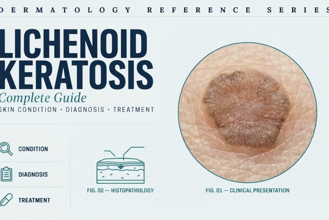
Lichenoid keratosis (LK) is a benign, non-cancerous skin growth that typically appears as a single, small, scaly patch on sun-exposed areas of the body, most often the chest, arms, or upper back. It’s sometimes called “lichen planus-like keratosis” because under the microscope, it resembles a skin condition called lichen planus, even though the two are not the same disease.
Most dermatologists believe lichenoid keratosis develops from an existing benign lesion, such as a solar lentigo (sun spot) or seborrheic keratosis, that undergoes an inflammatory, immune-driven change over time. In simple terms, your body’s immune system starts attacking a harmless skin spot, causing it to become inflamed, scaly, and sometimes itchy.
Key Facts at a Glance
- Type: Benign inflammatory skin lesion
- Common age group: Adults over 40, though it can appear earlier
- Common locations: Chest, back, forearms, and sun-exposed skin
- Color: Reddish-brown, violet, gray, or dark brown
- Size: Usually under 1 cm, though some grow larger
- Cause: Often linked to an inflammatory reaction within a pre-existing sun spot or seborrheic keratosis
What Causes Lichenoid Keratosis?
The exact trigger isn’t fully understood, but researchers and dermatologists point to a few contributing factors:
1. Sun Exposure
Long-term ultraviolet (UV) exposure is one of the biggest risk factors. Most lesions appear on skin that has had years of cumulative sun exposure.
2. Pre-Existing Skin Lesions
Many cases start as a solar lentigo or seborrheic keratosis that later becomes inflamed due to immune activity.
3. Immune Response
The body’s T-cells (a type of white blood cell) may mistakenly attack the affected skin cells, triggering the lichenoid (lichen-like) inflammatory pattern seen under the microscope.
4. Age-Related Skin Changes
As skin ages, it becomes more prone to pigment irregularities and benign growths, increasing the likelihood of lichenoid change.
It’s important to note: lichenoid keratosis is not contagious and is not caused by poor hygiene, diet, or lifestyle habits.
Signs and Symptoms of Lichenoid Keratosis
Because lichenoid keratosis can resemble other skin conditions, recognizing its typical presentation is helpful before your dermatology visit.
Common symptoms include:
- A single, isolated patch (rarely multiple lesions)
- Reddish-brown, gray, or violet coloring
- Slightly raised, scaly, or rough texture
- Mild itching in some cases
- Occasional tenderness if irritated
- Slow growth over weeks to months
Unlike many other skin lesions, lichenoid keratosis usually appears suddenly and then stabilizes or slowly regresses over time.
Lichenoid Keratosis vs. Other Skin Conditions
Because it mimics several other skin issues, proper diagnosis matters. Here’s how it compares:
Lichenoid Keratosis vs. Basal Cell Carcinoma
Basal cell carcinoma (BCC) tends to be pearly, shiny, and may bleed easily. Lichenoid keratosis is usually scaly and stable in size, without ulceration.
Lichenoid Keratosis vs. Actinic Keratosis
Actinic keratosis (AK) is a precancerous lesion caused by sun damage, often rough and sandpaper-like. Lichenoid keratosis can look similar but is not classified as precancerous.
Lichenoid Keratosis vs. Lichen Planus
Lichen planus usually appears as multiple itchy, purple, flat-topped bumps, often on the wrists or ankles. Lichenoid keratosis is typically a single lesion, not widespread.
Lichenoid Keratosis vs. Melanoma
This comparison matters most. Melanoma can have irregular borders, multiple colors, and asymmetry. Because lichenoid keratosis can sometimes mimic melanoma visually, dermatologists often recommend a biopsy to rule it out definitively.
How Is Lichenoid Keratosis Diagnosed?
Diagnosis typically involves a combination of visual examination and, in many cases, a biopsy.
Step 1: Clinical Examination
A dermatologist will examine the lesion’s color, shape, border, and texture using a dermatoscope, a specialized magnifying tool.
Step 2: Dermoscopy
Dermoscopy helps identify specific patterns, such as pigment structures or blood vessel arrangements, that distinguish lichenoid keratosis from cancerous lesions.
Step 3: Skin Biopsy
Because lichenoid keratosis can resemble melanoma or basal cell carcinoma, a biopsy is often recommended for confirmation. During a biopsy, a small tissue sample is examined under a microscope.
Histopathology (microscopic examination) typically shows:
- A band-like inflammatory infiltrate
- Damage to the basal cell layer
- Remnants of the original benign lesion (like a solar lentigo)
This microscopic pattern confirms the diagnosis and rules out malignancy.
Is Lichenoid Keratosis Dangerous?
No. Lichenoid keratosis is classified as a benign skin condition. It does not turn into cancer and does not spread to other parts of the body.
However, doctors recommend biopsy confirmation because:
- It can visually resemble melanoma or basal cell carcinoma
- Early skin cancers can sometimes mimic benign lesions
- Peace of mind matters for long-term skin monitoring
Once biopsy-confirmed, no further concern is typically needed, though your dermatologist may recommend routine skin checks.
Treatment Options for Lichenoid Keratosis
Treatment isn’t always necessary, especially once melanoma and other serious conditions are ruled out. However, options exist for cosmetic removal or symptom relief.
1. Watchful Waiting
Many lichenoid keratosis lesions fade or flatten on their own over several months. If the biopsy confirms it’s benign, doctors often recommend simply monitoring it.
2. Cryotherapy (Freezing)
Liquid nitrogen can be used to freeze and remove the lesion, similar to how skin tags or warts are treated.
3. Topical Corticosteroids
If the lesion is itchy or inflamed, a mild steroid cream may reduce irritation and redness.
4. Laser Therapy
For cosmetic concerns, laser treatment can lighten or remove the pigmented area with minimal scarring.
5. Surgical Excision
In rare cases, especially where diagnosis remains uncertain, a small surgical excision may be performed for both treatment and further pathological testing.
Which Treatment Is Best?
Treatment choice depends on:
- Lesion size and location
- Whether it’s causing discomfort
- Cosmetic preference
- Biopsy results
Your dermatologist will guide the most appropriate option based on your specific case.
Can Lichenoid Keratosis Be Prevented?
While you can’t fully prevent it, you can reduce your risk with these steps:
- Apply broad-spectrum sunscreen (SPF 30+) daily
- Wear protective clothing and hats during sun exposure
- Avoid tanning beds
- Get regular skin checks, especially after age 40
- Monitor existing sun spots or seborrheic keratoses for changes
Since sun damage is a major contributing factor, consistent sun protection is the most effective preventive strategy.
When Should You See a Dermatologist?
You should schedule a dermatology visit if you notice:
- A new spot that appears suddenly
- Changes in color, size, or shape of an existing spot
- Itching, bleeding, or tenderness
- Irregular borders or multiple colors within one lesion
Early evaluation ensures accurate diagnosis and rules out more serious skin conditions.
Frequently Asked Questions (FAQs)
1. Is lichenoid keratosis the same as skin cancer?
No. Lichenoid keratosis is a benign condition. It is not cancerous, but a biopsy is often done to rule out cancer because it can look similar to certain skin cancers.
2. Does lichenoid keratosis go away on its own?
Yes, in many cases, it can fade, flatten, or regress over several months without treatment.
3. Is lichenoid keratosis itchy?
It can be mildly itchy in some cases, though many lesions cause no symptoms at all.
4. Can lichenoid keratosis come back after removal?
Recurrence is uncommon, especially after cryotherapy, laser treatment, or excision.
5. Does insurance cover lichenoid keratosis treatment?
If a biopsy is medically necessary to rule out cancer, it is typically covered. Purely cosmetic removal may not be covered, depending on your insurance provider.
6. What does lichenoid keratosis look like on skin?
It usually appears as a single, scaly, reddish-brown or violet patch, often under 1 cm in size, located on sun-exposed skin.
7. Who is most at risk for lichenoid keratosis?
Adults over 40 with a history of sun exposure, fair skin, or existing sun spots and seborrheic keratoses are most commonly affected.
Conclusion
Lichenoid keratosis is a common, benign skin condition that often develops from long-term sun exposure and inflammatory changes within an existing skin spot. While it can resemble more serious conditions like melanoma or basal cell carcinoma, a proper dermatological examination and biopsy can confirm the diagnosis and rule out cancer.
Most cases require no aggressive treatment beyond monitoring, though options like cryotherapy, topical steroids, or laser therapy are available for symptom relief or cosmetic removal. If you notice a new or changing skin lesion, don’t self-diagnose. Schedule a dermatology appointment for accurate evaluation and peace of mind