
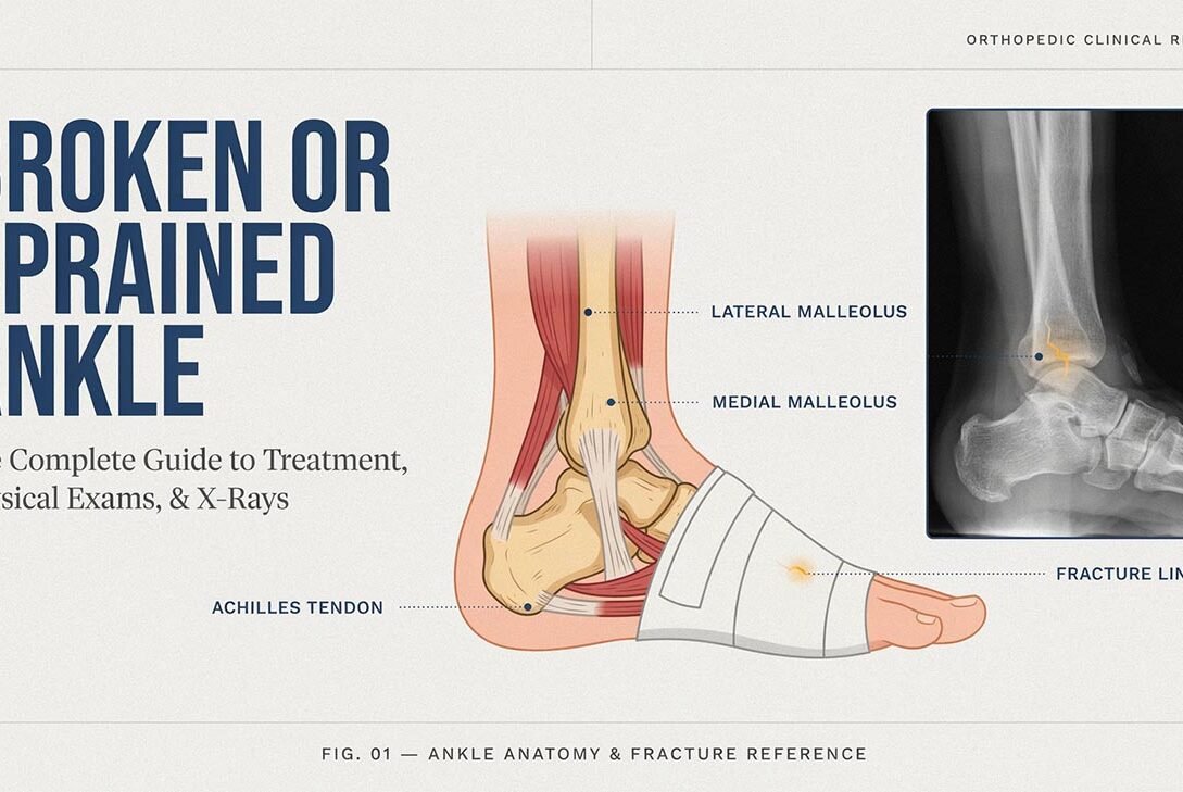
You rolled your ankle. Now it’s swelling, it hurts to walk, and you’re stuck googling at midnight trying to figure out whether you need a cast or just an ice pack. You’re not alone — ankle injuries are among the most common reasons people show up in urgent care and emergency rooms every single day.
The tricky part? A bad sprain and a fracture can feel almost identical in the first hour. Swelling, pain, and an unsteady walk show up with either injury, making them hard to tell apart in the moment. But how they’re treated — and how long they take to heal — can be very different. This guide walks you through how doctors actually tell the two apart, what the physical exam looks for, when X-rays are necessary, and what treatment looks like for each.
Broken vs. Sprained Ankle: What Makes Them Different?
A sprained ankle happens when the ligaments — the tough bands of tissue connecting bone to bone — get stretched or torn. This usually occurs when the foot twists or rolls beyond its normal range of motion.
A broken ankle (ankle fracture) means one or more of the bones in the ankle joint — the tibia, fibula, or talus — has actually cracked or split.
Here’s the confusing part: the mechanism of injury is often identical. You step off a curb wrong, land awkwardly during sport, or trip on uneven ground. Whether that force tears a ligament or snaps a bone depends on factors like the angle of the twist, the speed of impact, and even your bone density.
Quick comparison:
- Sprain — ligament injury, swelling around the joint, pain worsens with movement, usually no visible deformity
- Fracture — involves the bone rather than soft tissue; pain tends to be sharp and centered in one exact spot, and some people hear or feel a crack or pop when it happens
- Both — swelling, bruising, difficulty bearing weight, tenderness to touch
Common Causes of Ankle Injuries
Understanding what typically causes these injuries helps explain why they’re so common:
- A sudden inward or outward roll of the foot, common in sports involving quick direction changes — basketball, soccer, running
- Missing a step or stumbling on stairs
- Walking on uneven terrain or in poor lighting
- Wearing unsupportive footwear, especially high heels
- Landing awkwardly after a jump
- Motor vehicle accidents (usually higher-impact fractures)
- Falls from height
Athletes and physically active people face higher risk, but honestly, a simple misstep on a sidewalk causes just as many ankle injuries as sports do.
Signs and Symptoms: How to Tell What You’re Dealing With
While only imaging can confirm a diagnosis, certain symptoms point more strongly toward a fracture rather than a sprain.
Symptoms More Common with Sprains
- Swelling develops gradually over hours
- Pain is diffuse around the joint rather than at one exact point
- You can put some weight on the foot, even if it hurts
- Bruising appears but isn’t immediate
- Ankle feels “wobbly” or unstable rather than sharply painful
Symptoms More Common with Fractures
- Immediate, intense pain at a specific point on the bone
- The joint appears visibly misaligned or shifted out of its normal position
- Inability to bear any weight at all
- Swelling and bruising appear quickly, sometimes within minutes
- A sudden popping or snapping feeling that occurs as the ankle twists or impacts
- A loss of normal sensation in the foot, sometimes accompanied by a tingling or “asleep” feeling
When in doubt, err on the side of caution — visible deformity, zero weight-bearing ability, or severe pain should be treated as a likely fracture until you’re examined
The Physical Exam: What a Doctor Actually Checks
A physical examination for an ankle injury generally follows a step-by-step approach to identify the type and severity of the injury.
1. Inspection
The provider looks at the ankle for swelling, bruising, and any visible deformity. They’ll also compare it to your uninjured ankle for reference.
2. Palpation (Touch Test)
This is where the doctor gently presses along specific bony landmarks — particularly the back edge and tip of both the tibia (shin bone) and fibula (outer ankle bone), as extending to the base of the fifth metatarsal on the outer portion of the foot. Pain at these exact points is a red flag for fracture.
3. Range of Motion
You’ll be asked to move your ankle up, down, and side to side. Sharp, localized pain during this test often points toward a ligament injury, while inability to move the joint at all can suggest fracture.
4. Weight-Bearing Test
This examination often plays the most significant role in confirming the diagnosis. If you can take four steps without significant pain, it lowers the likelihood of a fracture requiring imaging.
5. Ottawa Ankle Rules
To avoid unnecessary imaging, emergency doctors often use this proven clinical decision tool to assess the need for an X-ray. It’s a well-validated, widely used framework that helps avoid unnecessary radiation exposure while still catching real fractures.
The Ottawa Ankle Rules: When You Actually Need an X-Ray
Not every twisted ankle needs imaging. The Ottawa Ankle Rules recommend an X-ray if any of the following are true:
- Bone tenderness at the back edge or tip of the lateral malleolus (outer ankle bone)
- Bone tenderness at the back edge or tip of the medial malleolus (inner ankle bone)
- Inability to bear weight both immediately after the injury and in the exam room (four steps)
- Bone tenderness at the base of the fifth metatarsal (for suspected foot injuries)
- Bone tenderness at the navicular bone (midfoot)
Because these guidelines reliably exclude most fractures, patients who do not meet the criteria generally do not require an X-ray. It’s one reason doctors don’t just X-ray every ankle injury automatically — it saves time, cost, and radiation exposure without compromising safety.
The Strengths and Limits of X-Rays for Ankle Injuries
X-rays are excellent at detecting bone fractures. A radiologist or physician will typically order three standard views: anteroposterior (AP), lateral, and mortise views, which together give a full picture of the joint from multiple angles.
However, X-rays have a limitation: they don’t show ligament damage. A completely normal X-ray doesn’t rule out a severe sprain. This is why the physical exam matters just as much as imaging — a doctor is piecing together the full picture, not just relying on a scan.
For complex injuries or cases where the diagnosis is unclear, further imaging may be needed to provide a more detailed evaluation
- MRI — best for detailed ligament, tendon, and cartilage assessment
- CT scan — useful for complex fractures involving the joint surface
- Stress X-rays — sometimes used to test joint stability under gentle pressure
Treatment: Sprain vs Fracture
Treating a Sprained Ankle
Most sprains, even moderate ones, respond well to conservative treatment using the well-known RICE method:
- Rest — give the joint a break from activities that put strain on it
- Use cold therapy for 15–20 minutes every few hours throughout the first two days to help reduce pain and swelling.
- Compression — an elastic bandage to reduce swelling
- Elevation — keeping the ankle above heart level when possible
Depending on severity, sprains are graded:
- Grade 1 (mild) — slight stretching, minimal instability, usually heals in 1–2 weeks
- Grade 2 (moderate) — partial tear, some joint looseness, healing takes 3–6 weeks
- Grade 3 (severe) — complete ligament tear, significant instability, may take several months and occasionally requires bracing or physical therapy
Physical therapy is often recommended for grade 2 and 3 sprains to restore strength, balance, and prevent future re-injury — ankle sprains have a notably high recurrence rate if rehab is skipped.
Treating a Broken Ankle
Fracture management is determined primarily by the break’s severity, location, and overall stability.
- Stable, non-displaced fractures — usually managed with a walking boot or cast for 4–6 weeks
- Displaced or unstable fractures — often require surgery to realign the bone with plates, screws, or rods
- Severe fractures involving the joint — may need longer immobilization and extended physical therapy afterward
Recovery timelines vary widely — anywhere from six weeks for a simple fracture to several months for surgical cases. Follow-up X-rays are typically taken during healing to confirm the bone is aligning properly.
When to Seek Emergency Care
Go to an emergency room immediately if you experience:
- Visible deformity of the ankle or foot
- Bone protruding through the skin
- Numbness, tingling, or a cold, pale foot (possible circulation issue)
- Complete inability to bear any weight
- Severe, unrelenting pain that doesn’t improve with rest
These signs suggest a more serious injury requiring urgent evaluation and possibly surgical intervention.
Frequently Asked Questions
1. Can you walk on a broken ankle?
Sometimes, yes — particularly with small, stable fractures. This is exactly why the Ottawa Ankle Rules and a proper physical exam matter; being able to walk doesn’t automatically rule out a fracture.
2. How long does a sprained ankle take to heal?
Mild sprains typically heal in 1–2 weeks, moderate sprains in 3–6 weeks, and severe sprains can take 2–3 months with proper rehabilitation.
3. Do all ankle injuries need an X-ray?
No. Doctors use clinical criteria like the Ottawa Ankle Rules to determine whether imaging is actually necessary, avoiding unnecessary X-rays when the exam findings are reassuring.
4. Sprain vs. Strain: A Simple Guide to Telling Them Apart?
A sprain affects ligaments (connecting bone to bone), while a strain affects muscles or tendons (connecting muscle to bone). Ankle injuries from rolling or twisting are almost always sprains, not strains.
5. Can a sprained ankle be worse than a fracture?
Yes, in some cases. A severe grade 3 sprain with complete ligament rupture can take longer to heal and cause more long-term instability than a small, stable fracture.
6. Should I use heat or ice on a sprained ankle?
Ice the injured area regularly throughout the first 48–72 hours to control inflammation and swelling. Heat can be introduced later to help with stiffness and circulation, but not in the acute swelling phase.
7. When can I return to sports after an ankle sprain?
This depends on severity, but most people can gradually return after 2–6 weeks, provided they’ve regained full range of motion, strength, and balance — ideally confirmed by a physical therapist or physician.
Conclusion
Telling a sprained ankle apart from a broken one isn’t always obvious just by looking at it — swelling, bruising, and pain can look similar for both. That’s exactly why the physical exam, guided by tools like the Ottawa Ankle Rules, plays such a central role in deciding whether an X-ray is actually needed. Sprains are treated with rest, ice, compression, and elevation, while fractures may need immobilization or surgery depending on severity. If you’re ever unsure, especially with visible deformity, inability to bear weight, or numbness, don’t wait it out — get it evaluated. A proper diagnosis early on makes a real difference in how quickly and completely your ankle heals.